



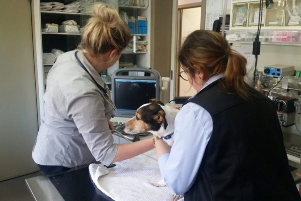
If your vet has just told you your dog or cat needs an X-ray or an ultrasound, it’s completely normal to feel a bit anxious. You might be wondering what’s actually involved, whether it’ll hurt, or whether your pet needs to be knocked out for it. The good news? These procedures are far more routine and far less stressful than most people expect.
Australia has one of the highest pet ownership rates in the world, with approximately 69% of households owning a pet. RSPCA Knowledgebase: and with so many of us sharing our lives with dogs and cats, veterinary diagnostic imaging has become a cornerstone of modern animal care.
This guide walks you through what to expect when your pet is referred for imaging at Keysborough Veterinary Practice.
What Is Veterinary Diagnostic Imaging?
Veterinary diagnostic imaging is the umbrella term for the tools vets use to see inside your pet’s body without making a single cut. The two most common are radiographs, often called X-rays, and ultrasound scans. Together, they give your vet a window into what’s going on beneath the surface, whether that’s a broken bone, a swollen organ, or something your pet swallowed that definitely wasn’t food.
Australia’s veterinary imaging market is projected to grow significantly, driven by increased pet ownership and growing awareness around animal health. Credence Research. That’s a reflection of something most pet owners already know instinctively: when your animal is unwell, you want answers, and you want them fast.
Why Vets Use X-Rays (Radiograph/Radiography)
A radiograph works by passing a small, controlled amount of radiation through your pet’s body to produce an image. Dense structures like bone absorb the radiation and show up white on the image, while softer areas appear in shades of grey. It’s the same basic principle used in human medicine, just scaled for animals.
Vets commonly use X-rays to assess:
- Broken or fractured bones: especially common in active dogs who love a run at the park
- Joint conditions like arthritis or hip dysplasia
- Foreign objects swallowed by curious pets (yes, socks and toy parts do show up)
- Chest or abdomen concerns: checking the lungs or organs for signs of disease
- Dental problems: Dental radiography is one of the most useful diagnostic tools in small animal care
- Spine and skeletal structure: important for pets showing signs of pain or limping
Radiography tends to be the first port of call because it’s quick, widely available, and excellent at showing bone and structure. If your dog has come home from Edithvale Reserve or Bangholme Trail favouring a leg, an X-ray is usually the first step your vet will take.
Radiography systems led veterinary imaging with the largest revenue share, reflecting their essential role in routine diagnostics. Mordor Intelligence
Why Vets Use Ultrasound
While an X-ray is brilliant for bones, it doesn’t tell the full story when it comes to soft tissue. That’s where ultrasound comes in. Ultrasound scanning is a painless, non-invasive procedure that uses sound waves to create real-time images of your pet’s internal organs.
A small handheld device called a transducer is pressed gently against your pet’s skin (usually after a little patch of fur is shaved and some gel is applied). The transducer sends sound waves into the body, which bounce back off internal organs to create a moving image on a screen.
Your vet might recommend an ultrasound to assess:
- Abdominal organs: including the liver, kidneys, spleen, and bladder
- The heart (an echocardiogram): to check for fluid, valve problems, or structural issues
- Pregnancy: to confirm and monitor a litter
- Masses or tumours: to better understand the size, location, and tissue involvement
- Abdominal pain of unknown origin
Unlike an X-ray, an ultrasound shows movement in real time, so your vet can actually watch the heart beating or the bowel moving. When it comes to evaluating soft tissue and internal organs, it’s often more informative than a radiograph alone.
Does My Pet Need Sedation?
This is one of the most common questions vets get asked, and the answer is: not always.
Many pets tolerate an X-ray perfectly well without any sedation. The procedure itself is quick, and if your pet is calm and reasonably cooperative, your vet can usually get the images they need with some gentle positioning and a reassuring hand.
That said, there are situations where a sedative may be needed:
- If your pet is in pain, positioning a sore limb for a radiograph can be uncomfortable, and a light sedative makes the process more humane
- If your pet is very anxious: a nervous dog or cat who won’t stay still makes it very difficult to get a clear, diagnostic-quality image
- For certain views, some radiograph angles are tricky to achieve without full relaxation
Having an ultrasound scan done is also generally very well tolerated. Most pets will simply lie still on a padded table while the transducer is moved across their belly. General anaesthetic for imaging alone is rare: it’s typically only needed for procedures like CT or MRI.
Your vet will always discuss sedation options with you beforehand and tailor the approach to your individual pet.
What Happens on the Day
Here’s a simple run-through of what to expect when you bring your pet in for imaging at Keysborough Veterinary Practice:
Arrival and check-in:
You’ll check in at the front desk. If sedation has been planned, you may have been asked to withhold food beforehand: your vet will have advised you on this. In many cases, patients are admitted into hospital for the procedure so the team can monitor them properly.
The imaging itself.
For an X-ray, your pet will be gently positioned on the table: sometimes on their side, sometimes on their back, depending on which area needs to be imaged. The process takes just a few minutes. For an ultrasound, a small area of fur is usually clipped, and gel is applied to the skin so the transducer can make good contact. You can think of a day to have an ultrasound as a very calm, hands-on check.
Reviewing the images.
Once the images are captured, your vet will review them: often while you wait, or within a short time after. Digital radiography means results are immediate. If the case is complex, images may be sent to a specialist radiologist for a second opinion.
Your hospital stay.
Whether your pet is admitted to the hospital for the day or discharged the same morning depends on the complexity of the case and whether sedation was involved. Your team will keep you informed throughout.
Understanding the Results
Once the images are reviewed, your vet will walk you through what they found in plain language. You’ll receive a clear diagnosis and treatment plan based on what the imaging shows. If further investigation is needed: bloodwork, specialist referral, or follow-up imaging, your vet will explain why and what the next steps look like.
Depending on what’s found, the treatment plan for your pet might be as straightforward as rest and medication, or it might involve further intervention such as orthopaedic surgery. Either way, imaging takes the guesswork out of the equation and means your vet can make decisions based on evidence, not just examination.
In urgent situations: say, your pet has swallowed something or taken a bad fall, imaging is often part of the urgent care process and may happen the same day you walk in.
On-Site Imaging at Keysborough Veterinary Practice
One of the biggest advantages of choosing a veterinary clinic like Keysi Vet on Chapel Road is that our hospital is fully equipped with on-site imaging. That means no referral delays, no stressful trips to an unfamiliar specialist clinic, and no waiting days for answers.
For dogs that spend a lot of time on Keysborough’s walking tracks and parks, musculoskeletal injuries are a real and recurring concern, and fast access to radiograph and ultrasound services means your vet can diagnose what’s going on and get a treatment plan moving quickly.
Our hospital is fully equipped to handle both routine imaging and more complex cases. Whether your pet needs a straightforward radiograph or a detailed ultrasound of their internal organs, the Keysi Vet team can often do it all in one visit.
Vets remain the most trusted source of advice for Australian pet owners, with 84% of dog owners and 69% of cat owners visiting a vet in the past year. GlobalPETS If you’re at all worried about your pet: whether it’s a limp, a lump, or something they shouldn’t have eaten: don’t wait. Book an appointment with us at X-rays and ultrasounds at Keysborough Veterinary Practice and let’s find out what’s going on.
Frequently Asked Questions
Is an X-ray safe for my pet?
Yes. Veterinary X-rays use very low levels of radiation, and the exposure from a single radiograph is minimal. The diagnostic benefit far outweighs any risk.
Will my pet be in pain during the scan?
Ultrasound scanning is a painless, non-invasive experience for your pet. X-rays are also painless: the only discomfort might come from positioning, which is managed with care and, if needed, sedation.
How long does imaging take?
Most X-ray and ultrasound appointments wrap up within 30–60 minutes, including prep time. If sedation is required, you may need to leave your pet with us for a few hours.
Can I be in the room?
In most cases, owners aren’t present in the X-ray room due to radiation safety protocols. However, your team will keep you updated, and you’ll typically be able to see your pet straight after.
Do I need a referral?
No referral needed. Contact Keysborough Veterinary Practice directly to book an appointment.
